Peer Reviewed
ECG education
Right bundle branch block
Recent articles on:
Hypertension
Hypertension
Abstract
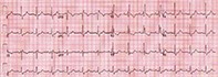
Amos is 80 years old and unknown to you. He has been to your practice a couple of times in the past three years. He takes no medications and tells you he has always had good health. He brings a medical report form for his driver’s licence and says his usual doctor has retired suddenly because of ill health. Amos shows you some normal blood test results from the past year and you ask him to tell you a bit about himself. As part of the driving medical report, you take his blood pressure, which is 160/95 mmHg. You arrange an ECG for Amos (see Figure).
Key Points
- In right bundle branch block, the QRS is over 120 msec, there is an ‘M’-shaped QRS complex in the leads V1 to V3 (also known as an RSR’ pattern) and slurring and widening of the S wave in the lateral limb leads I and aVL and the praecordial leads V5 and V6.
- Right bundle branch block may be considered normal if the cardiac echocardiogram is normal and there are no medical conditions to account for it.
- Right bundle branch block is common in children and is rarely of consequence.
- In people with incomplete right bundle branch block the QRS complex duration by definition will be less than 120 msec.
- Incomplete right bundle branch block is common and usually a normal finding but may be associated with underlying pathology.
- ST segments can still be interpreted with right bundle branch block (but not left bundle branch block) for the diagnosis of myocardial infarction.
Purchase the PDF version of this article
Already a subscriber? Login here.