Recurrent atrial fibrillation in a 73-year-old woman
Dr Miller is a GP in Sydney, a medical journalist and author, and the Medical Editor of Cardiology Today.
Articles in this section are inspired by, but not based on, real cases to illustrate the importance of knowledge about ECGs in relation to clinical situations in general practice. Management is not discussed in detail.
- The most common associations with atrial fibrillation (AF) include hypertensive heart disease, heart failure, valvular heart disease, ischaemic heart disease, obstructive sleep apnoea, obesity, excessive alcohol consumption, thyrotoxicosis and many lung diseases.
- The CHA2DS2-VASc score utilises the following risk factors, specifically in patients who have nonvalvular AF: – congestive heart failure – hypertension – age – diabetes – past stroke or transient ischaemic attack – vascular disease – female sex.
- If oral or intravenous medication (e.g. digoxin, calcium antagonists, beta blockers) to slow the heart rate pending spontaneous reversion; none of these medications control the rhythm in patients who have AF, just the rate.
- It is important to discuss with the patient in advance if they would prefer to use a rhythm or rate control strategy.
- Direct current or chemical cardioversion, aimed at rhythm control, is used for symptomatic patients and those with a reasonable prospect of maintaining sinus rhythm.
- Pulmonary vein isolation ablation is most frequently used in patients who fail to respond to medical therapy, in those who have contraindications or complications from oral antirrhythmic therapy, and in younger patients with no underlying cardiac disease and with a paroxysmal pattern of AF (as long-term success is higher).
Barbara is a mildly overweight 73-year-old woman who has been a patient of the practice for several years. She had breast cancer 10 years ago and has a mild cardiomyopathy, which is thought to be secondary to the chemotherapy agents she received. Eight weeks ago she had a second cardioversion for atrial fibrillation (AF), the first being two years previously. Barbara’s pulse during each occasion was an average 140 beats per minute, and she was haemodynamically stable on both occasions.
Barbara takes bisoprolol 2.5 mg daily, amiodarone 100 mg daily, frusemide 20 mg daily and perindopril 2.5 mg daily. She has not been able to tolerate calcium antagonists in the past, due to significant ankle swelling. She comes to see her GP for repeat prescriptions.
Q1. What are the important points for Barbara’s GP to cover in this consultation?
It is important for Barbara’s GP to ask if she has been aware of any heart palpitations, chest pain, shortness of breath or unusual light-headedness. Her pulse and blood pressure should be measured. If she is due for any routine tests, such as measurement of electrolyte levels, thyroid function tests or other relevant blood tests, they should be performed at this time.
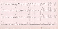
Q2. Barbara’s pulse rate is difficult to feel and her apex rate is irregularly irregular, fast and faint. She is asymptomatic apart from a modest reduction in exercise tolerance. Her blood pressure is 110/60 mmHg. Her GP performs an ECG (Figure). What does it show?
Barbara’s ECG shows AF at a rate of about 120 beats per minute. There are no ischaemic changes and no evidence of bundle branch blocks.
Q3. What is the immediate management of this patient?
Barbara’s cardiologist should be informed that she has relapsed into AF again. Barbara should see her cardiologist in the next few days. She should be advised that if she is feeling short of breath, faint, nauseated or has chest pain she needs to ring 000 to be hospitalised. Safety of driving should always be considered with patients with arrhythmias but Barbara has been consistently asymptomatic, so she should be safe to drive.
Q4. What is AF?
AF is a fast, irregularly irregular arrhythmia due to chaotic wavelets of electrical depolarisation continuously looping through the atria. No P waves are seen on the ECG, but fibrillatory waves are present and may range in amplitude from ‘coarse’ to ‘fine’ (less than 0.5 mm in amplitude and therefore more difficult to see). The ventricular rhythm is irregularly irregular and the rate is usually fast unless the patient is already on rate-slowing medication or there is atrioventricular (AV) node disease. The QRS complexes are normal in the absence of bundle branch blocks.
AF may arise from a focus of abnormal electrical activity (such as in the pulmonary veins) or from re-entry circuits arising from an abnormal (often dilated) left atrium. The most common association with AF include hypertensive heart disease, heart failure, valvular heart disease, ischaemic heart disease, obstructive sleep apnoea, obesity, excessive alcohol consumption, thyrotoxicosis and many lung diseases. It is the most common pathological arrhythmia and the incidence and prevalence both increase with age. There is a strong association with thromboembolism, due to the formation of thrombi from eddies and stasis within the left atrial appendage.
Q5. What is the CHA2DS2-VASc score?
The CHA2DS2-VASc score is a measure of risk assessment for stroke due to AF (in the absence of valvular heart disease). It is used to determine those patients who should be managed with new oral anticoagulant (NOAC) therapy or warfarin, and those for whom this is not recommended. Valvular AF has been excluded from the score because it carries a higher risk of thromboembolism, and anticoagulation with warfarin is indicated.
The CHA2DS2-VASc score uses the following risk factors, specifically in patients who have nonvalvular AF:
- congestive heart failure
- hypertension
- age 65 to 74 years
- age of 75 years or older
- diabetes
- past stroke or transient ischaemic attack
- vascular disease (i.e. coronary artery disease, peripheral artery disease or aortic atherosclerosis)
- female sex.
Each risk factor that is present is allocated one point, except for stroke or transient ischaemic attack, which is allocated two points. The annual risk of future stroke is then calculated as a percentage-adjusted stroke risk.
Men who have a CHA2DS2-VASc score of two or more or women with a CHA2DS2-VASc score of three or more should be started on an oral anticoagulant. Those with a CHA2DS2-VASc score of one (men) or two (women) or more are advised to consider an oral anticoagulant.
Patients with a CHA2DS2-VASc score of zero do not need long-term anticoagulation.
It should be noted that female sex will not be included in the soon to be published updated Australian AF guidelines for the CHA2DS2-VASc score (sexless CHA2DS2-VASc score). This will simplify recommendations by ensuring they are the same for both sexes.
Q6. What is pulmonary vein isolation ablation?
Ectopic beats can originate from the pulmonary vein and adjacent areas and trigger AF. Pulmonary vein isolation (PVI) radiofrequency ablation via catheterisation or surgery has been used, predominantly in patients with recurrent paroxysmal AF in whom it is highly successful for preventing recurrences. However, relapse is common and occurs in 30 to 50% of patients treated for AF. Nonpulmonary vein triggers account for about 20% of cases of relapse (e.g. arising in anatomical sites such as the superior vena cava or the posterior wall of the left atrium). Relapse is also more common after PVI ablation if there is underlying cardiac disease or the patient is older.
PVI ablation is most frequently used in patients who fail to respond to medical therapy, in those who have contraindications or complications from oral antirrhythmic therapy, and in younger patients with no underlying cardiac disease and with a paroxysmal pattern of AF (as long-term success is higher). Catheter ablation is also used in patients with atrial flutter and tends to be more successful.
Q7. How is acute AF managed?
Paroxysmal AF often terminates spontaneously within 24 hours and rate control may be useful during this time. Persistent/permanent AF does not revert spontaneously. Recurrent AF is defined as more than two episodes, as in Barbara’s case.
AF requires urgent management if the patient is haemodynamically unstable. This includes intravenous rate control (with medications such as digoxin, beta blockers, verapamil and amiodarone) and/or emergency direct current cardioversion.
If the patient is stable, they may be managed (often in the hospital setting) with oral or intravenous medication (e.g. digoxin, calcium antagonists, beta blockers) to slow the heart rate pending spontaneous reversion. Beta blockers may have some effect on rhythm control but the other medications do not control the rhythm in patients who have AF, just the rate. It is important to discuss with the patient in advance if they would prefer to use a rhythm or rate control strategy.
Direct current or chemical cardioversion, aimed at rhythm control, is used for symptomatic patients and those with a reasonable prospect of maintaining sinus rhythm, taking into account other factors. These include any underlying heart or lung conditions, the number of previous episodes of persistent AF they have experienced and the presence of a self-limiting or treatable precipitant for the current episode (such as an infective illness, injury, operation or alcohol binge). Direct current cardioversion has a higher success rate than chemical cardioversion; it is initially successful in more than 90% of such patients. Intravenous flecainide also has a very good cardioversion rate.
To minimise the risk of stroke or other thromboembolism, patients should generally be anticoagulated before cardioversion. They should also have left atrial thrombus ruled out by transoesophageal echocardiography if they have had more than 24 hours of AF within the preceding month when not therapeutically anticoagulated.
Q8. How do medications used to treat AF work?
Flecainide (a class IC antidysrhythmic drug) produces a dose-related decrease in cardiac conduction time throughout the atrial myocardium. It is contraindicated in patients with previous myocardial infarction or other significant structural heart disease.
Amiodarone (a class III antidysrhythmic drug that also has class I, II and IV actions) blocks sodium channels, calcium channels and alpha and beta adrenergic receptors and prolongs the myocardial refractory period. It may also inhibit sinus node function and AV conduction. It has multiple significant noncardiac side effects that are dependent on the dose used and duration of use.
Sotalol (a class III antidysrhythmic drug) is also a noncardiac-selective beta-adrenergic blocker (class II action). It prolongs the QT interval, especially in predisposed individuals or in combination with other medications.
Metoprolol, propranolol and atenolol (class II antidysrhythmic drugs) slow the atrial rate by blocking beta 1 adrenergic receptors in the myocardium. They slow AV conduction and therefore increase the block but are not as likely as other agents to terminate the arrhythmia.
Diltiazem and verapamil (class IV antidysrhythmic drugs) block calcium ion influx to cardiac smooth muscle and myocardium during depolarisation, therefore reducing heart rate via conduction through the AV node. These medications do not revert the AF to sinus rhythm.
Digoxin is now mainly used only in patients with both heart failure and supraventricular tachycardias, especially if other medications and catheter ablation are contraindicated. Digoxin is a positive inotrope that has a vagomimetic effect, decreasing conduction through the AV and sinus nodes. It is less effective if there is high sympathetic activity and does not revert to sinus rhythm.
Outcome